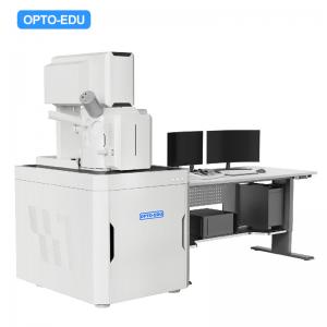
Add to Cart


| A63.7088 | |
| Resolution | 1.0nm@30KV(SE), 1.5nm@1KV(SE) |
| Magnification | 1x~2000000x |
| Electron Gun | Schottky Field Emission Gun |
| Voltage | Accelerating Voltage 0.02~30KV |
| Electron Beam | 1pA~20nA |
| Vacuum System | 1 Sputter Ion Pump, 1 Turbo Molecular Pump, 1 Mechanical Pump |
| Detector | SE in Lens, SE in Sample Room, BSE, CCD |
| Extend Port | Extend Ports On Sample Room For BSE, EDS, EBSD, CL etc. |
| Specimen Stage | 5 Axes Auto Stage, Travel Range: X=125mm, Y=125mm, Z=50mm, R=360°, T=-5°~+70° |
| Max Specimen | Specimen Room Dia.330mm, Height 260mm |
| Image System | Real Still Image Max Resolution 256x256~16k~16k Pixels |
| Computer & Software | PC Work Station Windows System, With Professional Image Analysis Software To Fully Control Whole SEM Microscope Operation, Mouse, Keyboard |
| Control Panel | Included |
| Dimension & Weight | Main Body 1900x1100x1800mm, Total Weight 800Kg |
| Optional Accessories | |
| A50.7091 | Ion Beam Cleaner |
| A50.7092 | Field Emission Gun Lamp |

| ▶ Superior Electron-optics Design ● Thermal field emission electron gun, stable beam, high imaging resolution ● Full tube acceleration technology ensures high imaging performance of electron beam at low acceleration voltage ● Composite lens design of electrostatic lens and magnetic lens, the objective lens has no magnetic leakage, and the imaging of magnetic samples is worry-free |

| ▶ Comprehensive Signal Collection System ● Can simultaneously collect signals from two types of secondary electrons, backscattered electrons and transmitted electrons. ● The sample morphology and composition contrast are displayed simultaneously to reveal the sample's microscopic morphology and composition information to the greatest extent. |


